The plants whose seeds have two cotyledons are called dicots. The structure of a dicot root varies greatly from that of the monocots. By understanding the structure of the dicot root and monocot root, we can make comparisons between them and distinguish them by studying them under a microscope.
Visit this page to learn about monocot root.
Anatomy of Dicot root (gram)
Dicot roots of gram shows the following distinct region in its Transverse section with the following features:
- Epiblema
- Cortex
- Endodermis
- Pericycle
- Vascular bundles
- Pith
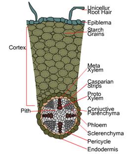
fig- T.S. of dicot root (gram shoeing its internal tissues organization
Different Regions of Dicot Root
1. Epiblema or Epidermis – It is the outermost unlayered with several unicellular root hairs. It consists of thin-walled, compactly arranged living parenchymatous cells. Usually, epiblema is characterized by the absence of stomata and cuticles. Sometimes the epiblema may be less cuticularised. It provides protection to the roots due to the presence of unicellular root hairs it also helps in the absorption of water and minerals from the soil.
2. Cortex – It is a thin-walled, multilayered region made from circular or polygonal parenchymatous cells. they usually have intercellular spaces. The cortical cells have no chloroplast but may contain leucoplast for storage of starch grains. The cortex is responsible for the transportation of water and salts from the root hairs to the center of the root.
3. Endodermis – It is the innermost layer of the cortex and covers the stele. It consists of compactly arranged barrel-shaped parenchyma without intercellular spaces. Most of the cells are characterized by the presence of special thickening of suberin and lignin on their radial and tangential walls called casparian strips. Some endodermal cell near the protoxylem has no casparian strips and are called passage cells or transfusion cells. These cells allow the radial diffusion of water and minerals through the endodermis.
4. Pericycle – It is the outermost layer of the stele and is composed of a uniseriate layer of parenchymatous cells without intercellular spaces. Some dicots and hydrophytes do not bear pericycle. Several lateral roots and lateral meristem arise from the pericycle region (hence lateral roots are endogenous in origin). At the time of secondary growth, it produces secondary cambium or phellogens.
5. Vascular bundles – They are 2-8 in number, radial, and arranged in a ring. Xylem and phloem bundles are separated from each other by parenchymatous cells called conjuctive or complementary tissue.
- Xylem is exarch (i.e. protoxylem towards the periphery and metaxylem towards the centre) and consists of tracheids, vessels, xylem parenchyma and xylem fibres.
- The pholem forms oval masses beneath the pericycle, alternating with xylem bundles. Pholem consists of sieve tubes, companion cells and pholem parenchyma. Usually pholem fibres are absent or reduced.
6. Pith – It is feebly developed and centrally located. It consists of thin-walled, polygonal parenchyma cells with intercellular spaces. In dicots roots, it may be reduced or absent. It helps in the storage of food materials.
Distinguishing Features of Dicot Root
The typical dicot roots show the following features.
- Epiblema is uniseriate, thin walled, colourless without intercellular spaces and produce unicellular root hairs, hence also called as piliferous layer or rhidodermis.
- Cortex is homogenous (without differentiation).
- Endodermis consists of barrel shaped compact parenchymatous cells. It contains both casparian stripes and passage cells.
- Pericycle uniseriate and become meristematic to give secondary roots and secondary tissues.
- Vascular bundles are radial; Xylem is exarch, number of xylem bundles varies from 2 to 4 rarely more (upto 6-8). Metaxylems are angular arranged in linear.
- Usually conjuctive tissues are well developed.
- Pith is very small or completed obliterated.
Recommended Readings