Based on the manner of orientation to the main axis of plant and direction of sunlight, leaves in angiosperms can be divided into two types
- Dorsi-ventral leaves
Dorsiventral leaves orient themselves at an angle to the main axis and perpendicular to the direction of sunlight. Most dicots have dorsi-ventral leaves that are net-veined, including most trees, bushes, garden plants and wildflowers. - Isobilateral leaves
Isobilateral leaves orient themselves parallel to the main axis and parallel to the direction of sunlight. Most monocots possess parallel-veined isobilateral leaves, including grasses and grasslike plants, lilies, irises, amaryllises etc.
Most leaves have certain common features like a covering of an epidermal layer on each surface. The ground tissue that occurs between the two epidermal layers is called mesophyll. Vascular bundles, commonly known as veins, are embedded in the mesophyll. The structure and characteristics of each of these layers differ greatly for dorsiventral and isobilateral leaves.
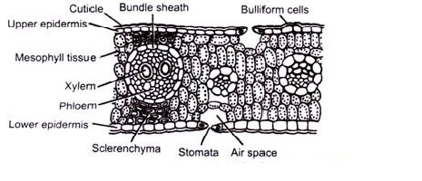
Anatomy of Leaf of maize (Zea mays)
We can study the anatomy of iso-bilateral leaf by taking a traverse section passing through the midrib region. The internal structure of isobilateral leaf of maize shows distinct layers of epidermis, mesophylls cells and vascular bundles with following features:

- Epidermis is single layered, present on both surfaces and has cuticles (cuticularized) and stomata on both surfaces (amphistomatic). It is composed of compactly arranged oval or barrel shaped thin walled parenchymatous cells.
A few cells in the Upper epidermis may become larger, less cuticularised; lens shaped and found on groups called motor cells or bulliform cells. These cells becomes empty and large and regulate the curling and uncurling (rolling up) of the leaves during dry conditions.
- Mesophyll is the ground tissue that is present between the two epidermal layers. It is not differentiated into palisade and spongy parenchyma and contains chloroplasts. It is composed of cells that are almost spherical, oval or angular with irregular intercellular spaces.
Mesophyll tissues are not found in the mid-vein region. In mid vein region, sclerenchymatous cells extend from the vascular bundle to the lower and upper epidermis. This extension of sclerenchyma is called bundle sheath extension.
- Vascular bundles are of two types- small bundles are abundant and larger bundles are found in between them. The bundles are conjoint, collateral, closed and each covered by parenchymatous bundle sheath cells containing starch grains.
Sclerenchymatous cells may be present on both sides of the large bundles.The larger bundles have distinct pholem towards the lower epidermis and xylem toward upper surface.
- Xylem consists of two pitted oval metaxylem; in between them, tracheids are also found. Xylem parenchyma are less numerous. Protoxylem is represented by a lysigenous cavity.
- Phloem has sieve tubes and companion cells. The small bundles are surrounded by individual sheaths and contain not distinct and less developed pholem and xylem.
Diagnostic Features of Isobilateral Leaf
- Two epidermal layers
- Amphistomatic – stomata on both layers
- Cuticularized – Cuticle is present on both epidermal layers
- Motor Cells – present in upper epidermis
- Undifferentialed mesophyll – tissue not differentiated into palisade and spongy parenchyma
- Bundle sheath – formed by sclerenchymatous cells that extend from the vascular bundle towards upper and lower epidermis
- Conjoint, collateral, closed – vascular bundles
- Two protoxylem and two xylem – present in each vascular bundle
- Hypodermal sclerenchyma – present on both sides of vascular bundle