The gram-positive cell wall is surrounded by a thick layer of peptidoglycan which acts as a protective layer for bacteria. And, during the Gram Stain test, the gram-positive cell wall turns purple (or violet), representing that the bacteria is of gram-positive type.
Bacteria with gram-positive cell walls are:
- Clostridium Botulinum
- Staphylococcus
- Streptococcus
- Enterococcus Faecalis
What is the structure of a gram-positive cell wall?
The structure of a gram-positive cell wall is mainly composed of a thick layer of Peptidoglycan, Periplasmic Space, Teichoic Acid, and Plasma Membrane.
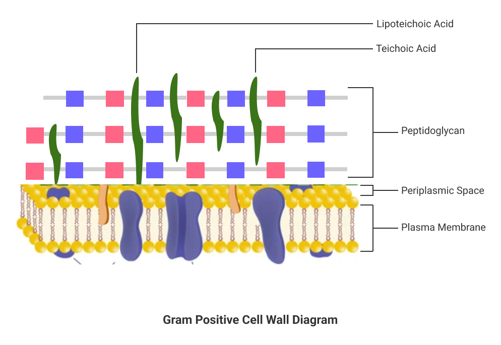
1. Peptidoglycan
The thick layer of peptidoglycan (about 20 nm – 80 nm) allows the gram-positive cell wall to absorb a lot of foreign materials. It also helps bacteria to survive in the host environment by decreasing the effect of antibiotics.
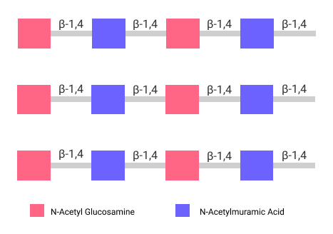
Peptidoglycan is an anionic polymer composed of N-Acetyl Glucosamine (NAG) and N-Acetyl Muramic Acid (NAM) linked with β-1,4 bonds.
2. Periplasmic Space
The indistinctive periplasmic space of the gram-positive cell wall is filled with Periplasm. Compared to the gram-negative cell wall, a relatively small number of proteins is present inside the Periplasm.
3. Teichoic Acids
Teichoic acids are also polymers composed of glycerol and ribitol linked by phosphate groups. They act as receptors for viruses that infect bacteria.
Sometimes the teichoic acid is attached to the plasma membrane (lipids), forming the lipoteichoic acid.
What are the characteristics of gram-positive cell walls?
Here’re some of the major characteristics of the gram-positive cell wall:
- The thickness of the peptidoglycan layer in a gram-positive cell wall is about 20 nm to 80 nm.
- During the staining procedure, the color of the cell wall cell is changed to violet.
- It consists of a cytoplasmic membrane beyond the peptidoglycan layer.
- The cell wall remains flexible to remodel during cell growth and cell division.
- It consists of teichoic and lipoteichoic acids that are helpful to recognize foreign bacteria.
- A gram-positive cell wall has a negative charge due to teichoic acids which have a COO – group.
How to identify gram-positive cell walls?
We use the Gram Staining Technique to identify the gram-positive cell wall. This technique was developed by Hans Christian Gram in 1884.
The gram staining technique involves coloring the bacterial cell wall. We can identify whether the cell wall is gram-positive or gram-negative based on the resulting color.
Gram Staining Test
The staining test involves 3 steps:
Step 1: Primary Staining
- Initially, we add crystal violet dye to the bacterial cell wall. It changes the color of the cell wall to purple.
- We then add iodine solution to form a crystal violet and iodine complex.
Step 2: Decolorization
- In this step, we wash the above complex using alcohol.
- Here, if the cell wall is of gram-positive type, it is found that the purple color of the cell wall remains as it is.
- If the cell wall is gram-negative, the color is completely washed.
Step 3: Counter Staining
- Now, we add a new color, safranin, to the sample.
- Since the gram-positive cell wall already has a purple color, the color remains, denoting that the cell wall is of gram-positive type.
- However, the gram-negative cell wall retains the pink color of safranin because Decolorization completely washes the purple color.
In this way, the cell wall that converts to purple after the staining test is a gram-positive cell wall.
Note: During the Decolorization step, the thick layer of Peptidoglycan prevents the purple color of the cell wall from washing off completely.
Gram-positive Vs. Gram-negative Cell Wall
Here’re some of the major differences between gram-positive and gram-negative cell walls.
| Gram-positive Cell Wall | Gram-negative Cell Wall |
| It has a thick layer of Peptidoglycan. | It has a thin layer of Peptidoglycan. |
| After the staining test, its color is changed to purple. | After the staining test, its color changed to pink. |
| It consists of a small number of proteins in Periplasm. | It consists of a large number of proteins in Periplasm. |