Mitosis cell division is the type of cell division cycle in which chromosomes in a cell nucleus are separated into two identical sets of chromosomes, each in its own nucleus.
The result of mitotic cell division is two daughter cells which are genetically identical to both each other and the parent cell. They resemble each other qualitatively as well as quantitatively. The mother and daughter have equal number of chromosomes (2n), and they are both haploid. Hence, this type of division is also called equational cell division.
In case of multicellular organisms, mitosis cell division ensures the increment in the number of cells as well as growth and development of the organisms. Mitosis cell division takes place in somatic or vegetative cells. Hence, it is also called vegetative cell division.
The process of mitosis is divided according to the development phases of the cells. It takes place as follows:
- Interphase
- Karyokinesis
- Cytokinesis
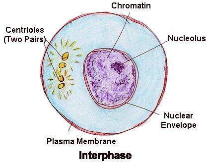
Interphase
Interphase is the time period between two successive cell divisions where the cell prepares itself for the process. It is classified as G1 (first gap), S (synthesis), and G2(second gap).
During these sub-phases, cell grows and volume increases by producing proteins and other cell organelles, nucleus stains darkly, chromosomes get duplicated, division takes place and various other biosynthesis takes place and the cell is metabolically very active. However, no visible changes are observed. Hence, this period is also known as “resting phase” of a cell.
Karyokinesis
Division of nucleus during the cell cycle known as karyokinesis. It brings about the division of nucleus to form two daughter nuclei. It takes place as follows:
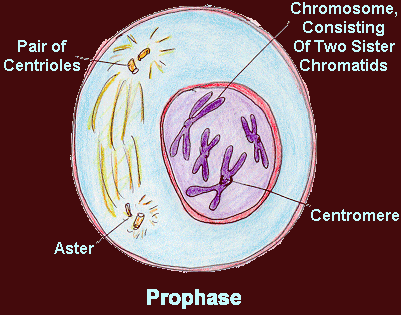
1. Prophase
This is the first stage of karyokinesis. During this period, the following changes appear in the cell:
- Nuclear membrane and nucleolus starts to disappear.
- The chromatin network of interphase starts to become shorter and thicker due to coiling as well as loss of water.
- Each chromosome is duplicated and is visible in the form of a pair of sister chromatids (pairs of identical copies of DNA joined at a point called the centromere) which remain attached at a common point called centrosome.
- By the late prophase, chromosomes are visible as thick rod-like structures and the nuclear membrane and nucleolus disappears completely.
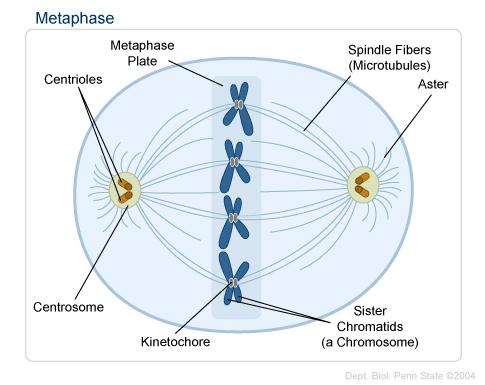
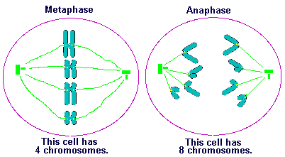
2. Metaphase
The following changes takes place during metaphase:
- Beginning of metaphase is characterized by the absence of nuclear membrane and nucleolus and the appearance of thread-like fiber called spindle fiber starting from the the poles of the cell.
- Chromosomes become shorter and thicker.
- Spindle fibers gets attached to the centromere of the chromosomes.
- All the chromosomes lie at the equatorial line of the cell. This process by which all the chromosomes at brought at the center of the cell is known as congression and the virtual plate where all the chromosomes lie is known as metaphasic plate.
3. Anaphase
The following changes takes place during anaphase:
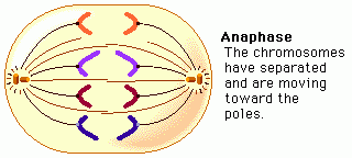
- During this period, sister chromatids are pulled towards opposite direction due to the contraction of spindle fiber.
- Due to the contraction of the spindle fiber, the centromere splits longitudinally resulting the single centromere for each chromatid, now called chromosomes. This is also supported by repulsion between two sister chromatids.
- Due to continuous contraction of spindle fiber, each chromosome now moves towards the opposite poles.
- By the late anaphase, spindle fiber disappears.
- During the pulling of the chromosomes by the spindle fiber, varies shapes of the chromosomes like (J) , (V) ,(i) , (l) are formed.
4. Telophase
Telophase is said to be the reversal of prophase.The following changes takes place in telophase:
- During the beginning of telophase, all chromosomes (which are already at the poles), now starts to uncoil or elongate. It is aided by the absorption of water.
- Slowly, nuclear membrane and nucleolus starts to appear.
- By the late telophase, nuclear membrane and nucleolus reappears and chromatin network starts to appear.
- At the end of telophase, two nuclei in a single cytoplasmic cell is formed.
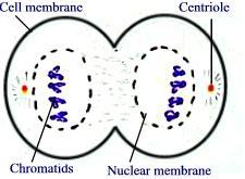
Cytokinesis
The division of cytoplasm and it’s contents is called cytokinesis. It takes place in plant cell and animal cell by cell plate method and call furrow method respectively. In case of plant cell, the remnants of spindle fiber organizes at the center of the cell. Similarly, vesicles from endoplasmic reticulum and golgi complex organizes at the middle of the cell to form solid plate-like structure called cell plate. Finally, cytoplasm divides between two newly formed nuclei and new daughter plant cells are formed. Whereas, in case of animal cell, constriction develops from peripheral side and it increases towards the center. When the constriction or furrow meets, the cytoplasm divides to give two daughter animal cells.
Significance of Mitotic cell division
- Mitotic cell division contributes to the growth of cells and development of multicellular organisms from unicellular organism.
- Old cells are replaced by new cells with the help of mitotic cell division.
- The lost body parts in some organisms is regenerated by mitotic cell division.
- It contributes to produce genetically same organisms by asexual reproduction (eg. Hydra).
- It helps in vegetative propagation of plants which is utilized in floriculture and horticulture.