The surface of our body and internal organs is subjected to wear, tear and injury. To protect the inner tissues, the outer lining of the body and organs is covered by a specialized group of cells called epithelium.
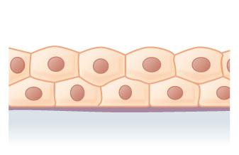
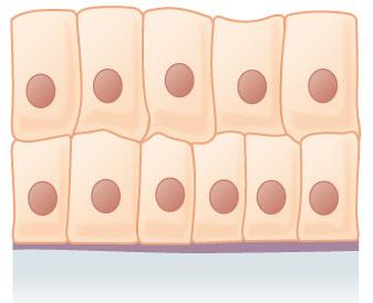
Epithelial tissues can contain a single layer of cells (Simple Epithelium) or many layers(Stratified Epithelium).
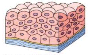
Stratified epithelial tissue is formed of several layers of epithelial cells of different shapes representing newly formed and mature cells. The superficial layer grows outward from below. Basement membranes are usually absent. Their main function is to protect underlying structure from mechanical wear and tear.
Types of Stratified Epithelium
Stratified Squamous Epithelium
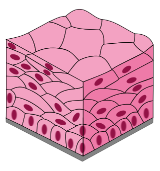
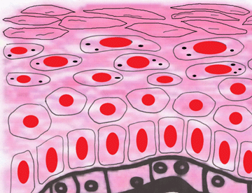
It is much thicker than simple epithelial tissue. This tissue is composed of several layers of cells of various shapes representing newly formed mature cells. The innermost layers mainly contains columnar cells cubiodal cells which are germinative in nature and as they grow towards surface. They become more flattened and then shed. It is two types: non-keratinized epithelium and keratinized epithelium.
- Non-keratinized stratified epithelium- This tissue is formed lower cubiodal cells. It is found on wet surfaces that are subjected to considerable wear and tear and are protected from drying. They are found in lining of mouth cavity, tongue, pharynx, oesophagus and vagina.
- Keratinized stratified epithelium- This tissue is found on outer dry surfaces of body like skin, hair claws and nails. The outer surface layer consists of dead epithelial cells. This tissue contains Keratin, a highly insoluble fibrous protein with water-proofing qualities. This epithelium is also resistant to friction and bacterial invasion.

Source: “Oral mucosa” by Wiki-minor – Own work. Licensed under CC BY-SA 3.0 via Commons.
Stratified Cuboidal Epithelium
The outermost layer consists of cubiodal cells. The innermost layer consists of germinative cells. This tissue forms the lining of ducts of glands.
Location- It is found in lining of sweat glands, mammary glands, conjunctiva of eyes and female urethera.

Function- help move nutrients and increase absorption. Also protects against pathogens/bacteria
Stratified Columnar Epithelium
It is also uncommon in the body; usually the basal layer consists of shortened, irregularly polyhedral cells. Only the superficial cells are columnar in form.
Location- This tissue is found in lining of vasa deferentia, male urethra, trachea and bronchi, etc.

Function- It helps in protection and secretion.
Transitional Epithelium
This tissue is composed of several layer of large, round or pear shaped cells. The superficial cells are larger and more rounded while basal layer consists of cubiodal cells.
Location- This tissue is found in urinary bladder and ureter.

Function- It helps in expansion of the organ.
Comparison between Types of Stratified Epithelium
| Location | Cells | Nuclei | Functions | |
|---|---|---|---|---|
|
Squamous |
Non-keratinized : in lining of mouth cavity, tongue, pharynx, oesophagus and vagina. Keratinized : on outer dry surfaces of body like skin, hair claws and nails |
Squamous cells apically, but basal layers may vary from cuboidal to columnar |
centrally located |
protection |
|
Cuboidal |
lining of sweat glands, mammary glands, conjunctiva of eyes and female urethera |
two layers |
centrally located and spherical |
absorption, secretion |
|
Columnar |
lining of vasa deferentia, male urethra, trachea and bronchi |
single layer of columnar cells on several layers of cuboidal cells |
basal and oval |
protection, secretion |
|
Transitional |
occurs only in bladder, ureter and urethra |
vary depending on stretch – apical cells often large, round and bi-nucleated |
centrally located |
distension |