A transverse section (T.S.) of a monocot root shows, from outside to inside: epidermis with root hairs, cortex, endodermis, pericycle, many radial vascular bundles (more than six) with exarch xylem, a little conjunctive tissue, and a large pith. Maize (Zea mays) is the usual example.
Below is a labelled diagram, what each region does, how to draw it in an exam, and how it differs from a dicot root, plus practice questions with answers.
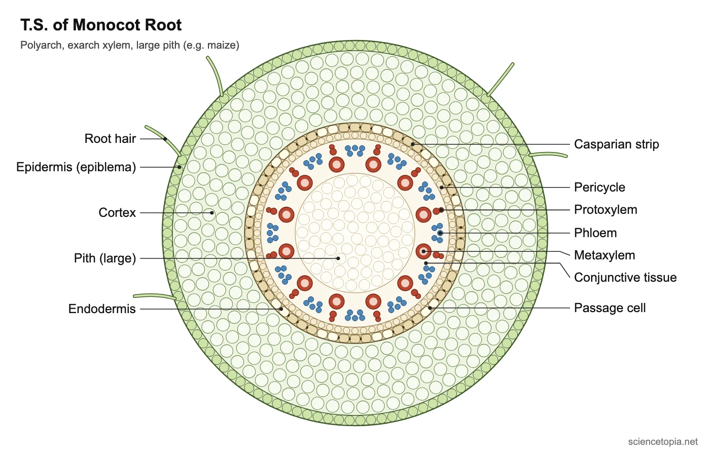
Regions of a monocot root and their functions
| Region | What it looks like | Function |
|---|---|---|
| Epidermis (epiblema) | Single layer of thin-walled cells, no cuticle. Many cells form unicellular root hairs. | Absorbs water and minerals; protects. |
| Cortex | Many layers of thin-walled parenchyma with intercellular spaces. In older roots the outer layers may become suberised (exodermis). | Stores food; passes water inward. |
| Endodermis | One layer of barrel-shaped cells with Casparian strips. Passage cells lie opposite the protoxylem. | Controls entry of water and minerals into the stele. |
| Pericycle | One layer of cells inside the endodermis. | Lateral roots arise here. It does not form cambium, because monocot roots have no secondary growth. |
| Vascular bundles | Radial, polyarch (usually more than 6 xylem patches), exarch xylem. Metaxylem vessels are rounded or oval. | Xylem carries water up; phloem carries food. |
| Conjunctive tissue | Narrow band between xylem and phloem; can be sclerenchymatous in older roots. | Separates xylem and phloem; adds strength. |
| Pith | Large and well developed, made of parenchyma. | Stores food. |
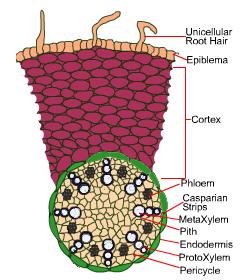
How to draw the T.S. of a monocot root (6 steps)
- Draw a large circle for the outline, a thin epidermis band and a few root hairs.
- Fill a wide band with rounded cortex cells (one sector is enough).
- Draw the endodermis as a ring of barrel-shaped cells with dots for Casparian strips, then a thin pericycle ring inside it.
- Draw a large circle in the centre for the pith, filled with big parenchyma cells.
- Around the pith, draw 8 to 12 xylem patches: a small protoxylem cell near the pericycle and one large round metaxylem vessel towards the pith.
- Put a small phloem patch between each pair of xylem patches, then label every region.
Examiners look for: many radial bundles, exarch xylem, rounded metaxylem and a large pith.
Monocot root vs dicot root
| Feature | Monocot root | Dicot root |
|---|---|---|
| Number of xylem patches | More than 6 (polyarch) | 2 to 4 (diarch to tetrarch), rarely up to 6 |
| Pith | Large and well developed | Small or absent |
| Metaxylem shape | Rounded or oval | Angular or polygonal |
| Pericycle forms | Lateral roots only | Lateral roots, vascular cambium and cork cambium |
| Secondary growth | No | Yes |
Practice questions
1. Why don’t monocot roots show secondary growth?
They have no cambium between the xylem and phloem, and the pericycle doesn’t form one, so no secondary xylem or phloem is added.
2. A T.S. shows 14 xylem patches, exarch xylem and a large pith. Identify it.
A monocot root. Polyarch bundles and a large pith are monocot root features.
3. What does exarch xylem mean in a root?
The protoxylem (first-formed xylem) lies towards the outside, near the pericycle, and the metaxylem towards the centre.
Frequently asked questions
What is the internal structure of a monocot root?
Epidermis with root hairs, a wide parenchymatous cortex, endodermis with Casparian strips, pericycle, many radial vascular bundles with exarch xylem, a little conjunctive tissue and a large pith.
How many vascular bundles are there in a monocot root?
Usually more than six xylem patches, alternating with as many phloem patches. That arrangement is called polyarch.
Are passage cells present in a monocot root?
Yes. Endodermal cells opposite the protoxylem stay thin-walled as passage cells, letting water move into the xylem.
Related